
CYTO Virtual Interactive 2021 - Scientific Tutorials
-
Register
- Visitor - $250
- Bronze Member - Free!
- Platinum Member - Free!
- Platinum - Free!
- Bronze Member 3-Year - Free!
- Silver Member 3-Year - Free!
- Platinum Member 3-Year - Free!
Products in this package:
- Virtual Training Strategies for an SRL
- Computational Cytometry – a basic guide for users and resources for SRLs
- Barking technology does not bite: Introduction to flow cytometry
- Image-Based High-Throughput Screening, High-Content Analysis and High-Throughput Profiling
- Size Matters: Practical Considerations When Starting a Small Particle Analysis Experiment
- Biosafety Principles for Flow Cytometry
- Immunophenotyping Best Practices – A Practical Guide for Experiment Planning, Panel Design and Required Controls
- Best Practices in Genomic Cytometry and Single Cell Multi-omics
-
Contains 3 Component(s), Includes Credits
A CYTO Virtual Interactive 2021 Oral Presentation presented by Shobana Stassen Keywords: Chimeric antigen receptors, CAR, surface receptor quantification
Overview
Background: Antigen density has emerged as a major factor determining the potency of CAR T cells. Traditional designs, such as KYMRIAH-based constructs have shown impressive clinical benefit when targeting B lineage antigens but manifest a high antigen density threshold for full activation, increasing the risk of relapse with antigen low variants, as observed following CARs targeting CD22 (Fry et al., 2018). In solid tumors, striking clinical results are lacking. Challenges include that truly tumor-specific antigens are rare and typically expressed at lower and more heterogeneous antigen densities. An accurate understanding of molecules/cell on tumor cells is crucial, as signal strength can be tuned by modifying CAR design towards an optimal therapeutic window and disease models used for CAR T cell development must be validated to express clinically relevant levels of target antigen, if they are to inform predicted potency against disease in humans.
Methods: Using flow cytometry to directly measure molecules/cell of immunotherapy-relevant targets including Glypican-2 (GPC2) on neuroblastoma cells in patients, we developed and applied a multicolor antigen density quantification assay on nine bone marrow samples infiltrated with metastatic tumor obtained and tested at 2 different institutions. Hematopoietic cells were excluded from the analysis based upon CD45, and bone marrow stromal cells based upon CD13 expression (Theodorakos et al., 2019). Gated CD45–CD13– cells co-expressed NCAM and GD2, known to be expressed on neuroblastoma (Warzynski et al., 2002) and were quantified in addition to GPC2, L1CAM and ALK. The fluorescence signal from the saturating antibody-fluorophore conjugates was converted into molecules/cell by using the reagent F/P ratio along with calibrated BD QuantiBRITETM beads and Custom Quantitation Beads containing known numbers of fluorophore molecules per bead.
Results: Metastatic neuroblastoma in the bone marrow demonstrated 4262±703 GPC2 molecules/cell, which is significantly lower than levels measured on the majority of neuroblastoma cell lines (p=0.0236). In comparison to several other immunotherapy targets we observed that GPC2 antigen density is lower than GD2, NCAM and L1CAM, but higher than anaplastic lymphoma kinase (ALK). The highest antigen density was observed for the disialoganglioside GD2, which demonstrated almost 30x more molecules than GPC2 and was expressed at significantly higher levels than the other targets tested. Levels of GPC2 on clinical specimens were approximating the level present on the neuroblastoma cell line SMS-SAN and found to be below the threshold required to control tumor growth for traditionally designed GPC2 CAR T cells. Sequential modifications to the CAR architecture, such as CD28-derived hinge-transmembrane and signaling domains and the overexpression of cJun resulted in a potent lead GPC2-CAR capable of impressive tumor regression mediating long-term complete responses in the absence of toxicity.
Conclusion: Our data emphasize that overexpressed tumor antigens can vary widely in the level and variation of expression and illustrate the profound impact of antigen density on the potency of CAR T cells. We highlight an essential role for the accurate quantitation of antigen density on clinical samples and show that selecting disease models for CAR T cell development informed by these measurements resulted in a promising lead candidate available for testing in early phase clinical trials.
Speaker

Shobana Stassen
Researcher
University of Hong KongShobana Stassen received her Bachelor of Science in Engineering (Hons) from Princeton University in 2010 and her Master of Science in Electrical and Electronic Engineering (Distinction) from the University of Hong Kong in 2019. She is currently a researcher at the Applied Life Photonics group at the University of Hong Kong. Her research interests are primarily in the development graph based statistical methods suitable for multi-omic and scalable single-cell analysis.
CMLE Credit: 1.0
-
Contains 3 Component(s), Includes Credits
A CYTO Virtual 2021 Scientific Tutorial presented by Swaine Chen, PhD, MD
About the Presenter
Swaine Chen, PhD, MD
Associate Professor, Department of Medicine
National University of Singapore
Group Leader, Infectious Diseases Group, Genome Institute of Singapore, A*STARCMLE Credit: 1.0
-
Contains 3 Component(s), Includes Credits
A CYTO Virtual Interactive 2021 Scientific Tutorial presented by Kathy Daniels, Rui Gardner, and Derek Davies Keywords: Training, training strategies, engagement, education
Overview
Education and training of researchers is one of the most important services provided by Shared Resource Laboratories (SRLs). As a response to the current global pandemic, many SRLs have been forced to shut down operations or enforce social distancing policies which have severely limited traditional training approaches. Education and training has been changing over the last few years and SRLs have had to adapt to different modes of delivering education, training, and on-going support. In this tutorial we will review workflows and best practices needed to successfully carry out remote theoretical and hands on training. This will include an overview of remote access tools, available technologies to implement these trainings, metrics that allow assessment of success, and best approaches to keep trainees engaged. We will also discuss the challenges that come with the transition to virtual training approaches, practical solutions to address them and how the training landscape might change in the coming years.
Speakers
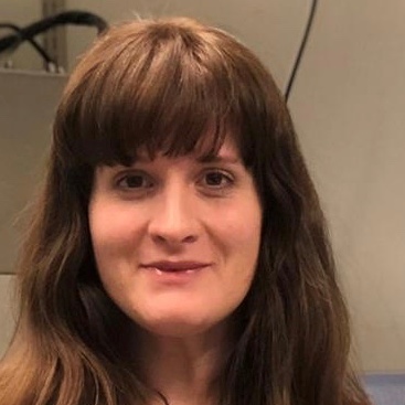
Kathy Daniels
Manager, Flow Cytometry Core Facility
Whitehead Institute
Cambridge, MAKathy currently serves as the manager of the Flow Cytometry Core Facility at the Whitehead Institute in Cambridge, MA. She has been working in flow cytometry for the past 8 years and is actively involved with the local and international cytometry community. Her goal is to empower researchers through a better understanding of the technology while providing increased open access educational resources to cytometrists worldwide. Kathy is currently the Vice President of MetroFlow and an ISAC SRL Emerging Leader.
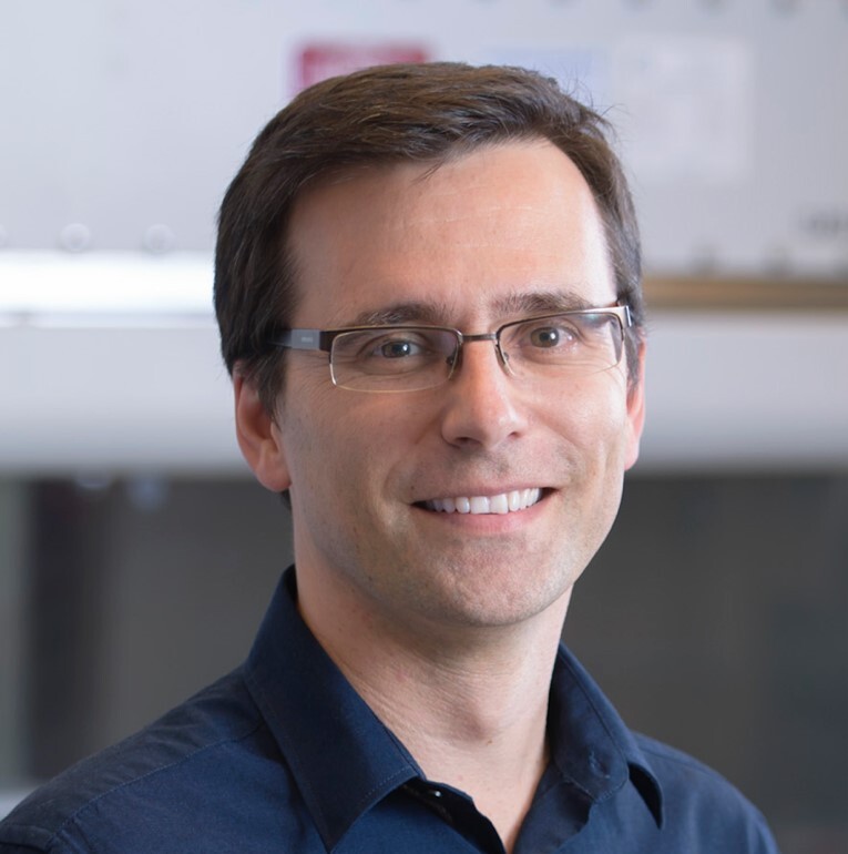
Rui Gardner, PhD
Lead, Flow Cytometry Core Facility
Memorial Sloan Kettering Cancer Center
New YorkRui started his career in science as a mathematical biologist, working in the lab of Dr. Michael Savageau at the University of Michigan, work that led to his PhD in Biomedicine in 2004 for the University of Porto in Portugal. In 2007, he became Head of the Flow Cytometry Core Facility at Instituto Gulbenkian de Ciência in Portugal. Serving more than 40 research groups in many diverse fields including immunology, inflammation, stem-cell biology, microbiology, virology, plant biology, among others, gave him a comprehensive experience in many flow cytometry techniques. In 2016 he was hired to lead the Flow Cytometry Core Facility at Memorial Sloan Kettering Cancer Center in New York. Passionate about science and technology, his background in mathematics, physics, and biology has allowed him to bridge the gap between the operational and technical details in flow cytometry and the science within flow applications. For the past 15 years, he has lectured countless courses in flow cytometry and cell sorting around the world, as well as been involved in organizing various international and regional workshops or scientific meetings on flow cytometry and core management both in Europe and the US. As councilor for the International Society for Advancement of Cytometry from 2012-2016 and the Iberian Cytometry Society (SIC) from 2015-2017, he has served on many committees and led the efforts to introduce the first ISAC SRL leadership programs that have benefited many young leaders of our community.
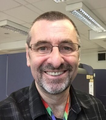
Derek Davies
STP Training Lead
Francis Crick Institute
London, UKDerek is the Science Technology Platform (STP) training lead at the Francis Crick Institute in London. He began his career in cytometry in 1980 using a microdensitometer to measure DNA content in cells, before moving into flow cytometry in 1985. For many years he ran the core flow facility at the London Research Institute and latterly the Francis Crick Institute looking after 30 cytometers and overseeing a staff of 12. In all that time, he has been actively involved in teaching on local, national, and international courses on all aspects of cytometry, including analysis and cell sorting. His current role is centered around standardizing training delivery within STPs and also developing training materials to develop competence and capability in cytometry and other technologies to the wider scientific community. He is chair of flowcytometryUK, sits on the Flow Cytometry Committee of the Royal Microscopical Society and is a former ISAC Councilor (2010-2014).
CMLE Credit: 1.0
-
Contains 3 Component(s), Includes Credits
A CYTO Virtual Interactive 2021 Scientific Tutorial presented by Sofie Van Gassen and Diana Bonilla-Escobar Keywords: computational analysis, basic
Overview
Cytometry data is typically analyzed by creating manual sequential gates in two-dimensional plots to find cell populations of interest. The recent development in instrumentation and reagents has dramatically increased the number of simultaneous parameters that can be measured in a single cell, leading to massive and complex datasets and the need for faster, scalable, and less subjective analytical solutions. This transformation in how data is being analyzed requires learning new skills and unsupervised computational analysis, which can be daunting and intimidating. The goal for this tutorial is to simplify the understanding of the tools, packages, and software available for computational data analysis in the SRL context.
Speakers
Sofie Van Gassen, PhD
Postdoctoral Researcher
VIB-UGent Center for Inflammation Research
Ghent, BelgiumSofie Van Gassen received her PhD in computer science engineering from Ghent University in 2017. During her PhD, she developed machine learning techniques for flow cytometry data, including the FlowSOM algorithm. She is now working on tools for computational cytometry in clinical studies during her postdoc in the Saeys lab (VIB-UGent Center for Inflammation Research). She is an FWO postdoctoral fellow and an ISAC Ingram Marylou Scholar.
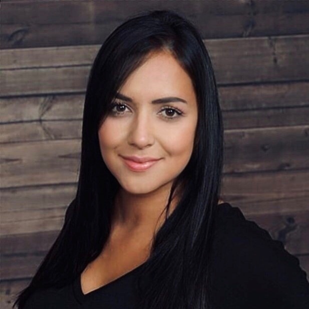
Diana Bonilla-Escobar, PhD
US Technical Lead for Application Support
Cytek Biosciences
Fremont, CADiana Bonilla Escobar is an immunologist with a PhD from Texas A&M University, a postdoctoral degree from Baylor College of Medicine, and an ASCP-accreditation as a cytometry specialist. She has more than 20 years of experience as a biomedical scientist for a variety of applications, including infectious diseases and cancer at MD Anderson Cancer Center. She is a former ISAC SRL Emerging Leader and highly involved in education with cytometry societies such as ISAC, FlowTex, and LatinFlow. She currently works as the US Technical Lead for Application Support at Cytek Biosciences.
CMLE Credit: 1.0
-
Contains 3 Component(s), Includes Credits
A CYTO Virtual Interactive 2021 Scientific Tutorial presented by Roxana del Rio-Guerra and Mariela Bollati-Fogolín Keywords: basic, introductory, cytometry applications
Overview
This tutorial will present the basic concepts of flow cytometry to encourage you to start using this technology. It will also provide simple and useful application examples that will allow you to sit in front of the cytometer and use it!
Speakers
Roxana del Rio-Guerra, PhD
Director of the Flow Cytometry and Cell Sorting Facility
Larner College of Medicine, University of Vermont
Burlington, VTAfter completing her PhD in immunology by the National Autonomous University of Mexico, Roxana was working as a postdoctoral associate in the Immunobiology Program at the University of Vermont (UVM) under the direction of Dr. Cory Teuscher. Roxana has more than 20 years of experiences in the use of flow cytometry within basic and applied immunology research. Roxana provides scientific and technical expertise in the design of flow cytometry experiments and data analysis, including immunophenotype, intracellular staining, cell cycle, proliferation, apoptosis, etc. Roxana has a passion for teaching this technology to the incoming generation of scientists.
Mariela Bollati-Fogolín, PhD
Head of the Cell Biology Unit
Institut Pasteur of Montevideo
UruguayMariela holds a PhD in biological sciences from the Universidad Nacional del Litoral, Argentina. She performed a postdoctoral training at the Helmholtz Centre for Infection Research in Germany, where she became passionate about flow cytometry and the strengths that this technology offers. At Institut Pasteur de Montevideo, she has driven the creation of a vibrant flow cytometry facility that supports and assists scientists and companies from throughout Latin America region. She has been engaged in the delivery of several flow cytometry training courses in the South America. She is actively involved in different activities related to public engagement as well as promoting women in science.
CMLE Credit: 1.0
-
Contains 3 Component(s), Includes Credits
A CYTO Virtual Interactive 2021 Scientific Tutorial presented by David Egan and Joshua Harrill Keywords: Image based high throughput screening, screening, informatics, data processing, multiplexed
Overview
This tutorial session introduces basic concepts of imaging-based high-throughput screening (HTS) and high-content analysis (HCA). Conventional HTS assays are designed to evaluate a discrete cellular process and produce a single or small number of quantitative outputs. In contrast, imaging-based HTS/HCA approaches measure dozens to thousands of features and provide highly multiplexed quantitative results. Topics covered in this tutorial include (but are not limited to) considerations for imaging equipment, biological model selection, endpoint selection, imaging assay design, identification and use of positive control and reference treatments, methods for evaluating assays’ dynamic ranges, and approaches for assessing assays’ reproducibility, as well as informatics and data processing method allowing design and interpretation of results. In particular, we will focus on the use of modern machine learning and artificial intelligence in HCA/HTS in the context of predictive toxicology. We will discuss how the technologies have been applied in various disease fields and the challenges associated with the implementation of these methods. The attendees familiar with multiparametric flow cytometry techniques will gain a basic foundational knowledge of quantitative image-based single-cell measurements and recognize the similarities and differences between the HCA approaches and flow cytometry.
In summary, this presentation's goals are the following:
- Provide an overview of the HTS and HCA methods
- Understand how the combination of HTS/HCA and artificial intelligence can enhance understanding of complex biological processes and enable predictive toxicology
- Gain insights into the challenges associated with the implementation of HTS/HCS methods.
Speakers
David Egan, PhD
Co-Founder and CEO
Core Life Analytics
Netherlands
Joshua Harrill, PhD
Lead Investigator
High-Throughput Transcriptomics
High-Throughput Phenotypic Profiling Hazard Screening
CCTECMLE Credit: 1.0
-
Contains 3 Component(s), Includes Credits
A CYTO Virtual Interactive 2021 Scientific Tutorial presented by Vera Tang and Gelo Dela Cruz Keywords: experimental design, extracellular vesicles, submicron, small particle, best practices
Overview
Would you like to know more about using flow cytometry to analyze biological particles that are smaller than cells? Many researchers are now looking into using flow cytometry for the phenotypic analysis of small particles such as extracellular vesicles (EVs), organelles, and viruses. The majority of this small particle research utilizes conventional flow cytometers that were designed for the analysis of cells, which are much larger in size with proportionately higher levels of antigen expression. Analysis of small particles using these instruments pushes them to the limit of detection. Therefore, having the right controls with best practices becomes very important for the interpretation and validation of the experimental data. Join us for this tutorial as we go over practical considerations, best practices, and current recommendations for analyzing small particles using conventional flow cytometers.
Speaker
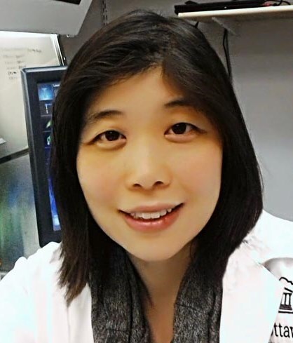
Vera Tang, PhD
Operations Manager|Adjunct Professor
Flow Cytometry & Virometry Core Facility
University of Ottawa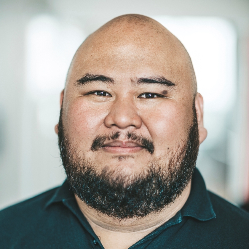
Gelo Dela Cruz, SCYM(ASCP)CM
Manager
Flow Cytometry Platform
NNF Center for Stem Cell Biology
DanStem
University of CopenhagenCMLE Credit: 1.0
-
Contains 3 Component(s), Includes Credits
A CYTO Virtual Interactive 2021 Scientific Tutorial presented by Kristen Reifel, Evan Jellison, Avrill Aspland, Iyadh Douagi, and Stephen Perfetto Keywords: General biosafety, risk assessment, infection agents, aerosol containment
Overview
This tutorial will provide a summary of biosafety principles as they apply to flow cytometry, including the general biosafety guidelines for cell sorting and considerations for cell analysis. Specifically, this tutorial will include guidance on performing risk assessments, provide recommendations for handling samples containing SARS-CoV-2 and other high risk group (BSL-2/3) infectious agents, and include a review of how to perform aerosol containment testing. We will also provide a forum to discuss specific scenarios that operators or core facility managers encounter with biosafety committee experts. After participation in this tutorial, the attendee will understand the principles and practices of biosafety as they pertain to flow cytometry.
Speakers
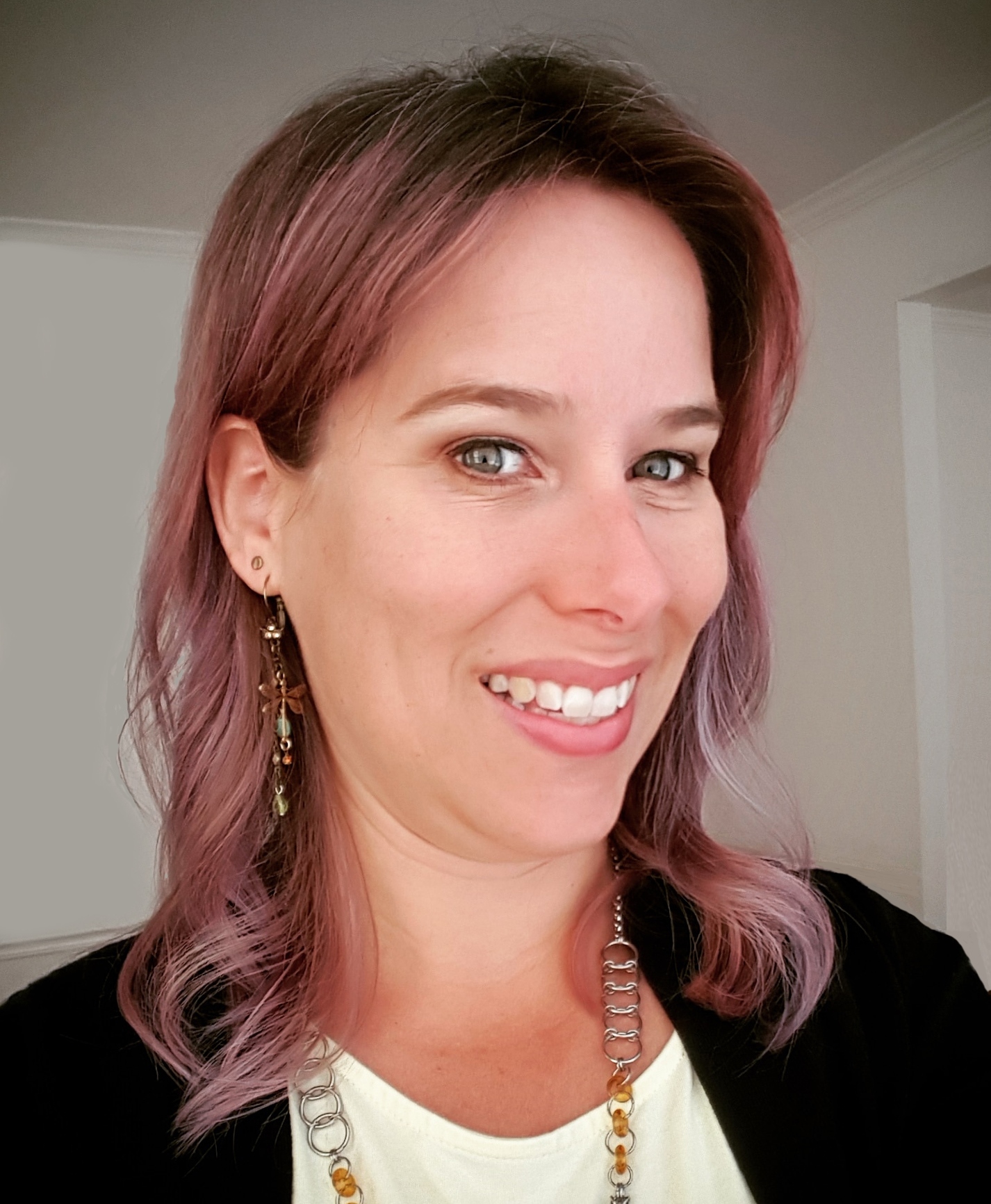
Kristen Reifel, PhD
Principal Investigator, Genomics
National Biodefense Analysis and Countermeasures Center
Frederick, MDDr. Kristen M. Reifel is a principal investigator in the Genomics Group at the National Biodefense Analysis and Countermeasures Center (NBACC) and runs a flow cytometry facility within BSL-2 and BSL-3 containment laboratories. She is also a member of the ISAC Biosafety Committee and is an ISAC SRL Emerging Leader. She received her BS in biology from UC Santa Barbara, MS in ecology from San Diego State University, and PhD in oceanography from the University of Southern California. Prior to joining NBACC, Dr. Reifel was a research associate at the J.J. MacIsaac Flow Cytometry Facility at the Bigelow Laboratory for Ocean Sciences and a postdoctoral fellow at Oregon State University. Dr. Reifel has used flow cytometry in research on both land and at sea, and she is now focused on single cell analysis and sorting of microbes from complex samples for genomic sequencing at NBACC.
Evan Jellison, PhD
Assistant Professor and Director of Flow Cytometry
UCONN School of MedicineEvan earned his doctoral degree from the University of Massachusetts Medical School, where he studied immune responses to viral infections. He then entered into postdoctoral training at UCONN School of Medicine studying immune tolerance in memory T cells. Throughout his training, flow cytometry played a vital role in Evan’s research and he gained substantial expertise in the technology. This ultimately resulted in him being hired to continue on at UCONN School of Medicine as an assistant professor and director of flow cytometry. Evan continues to play an important role in nearly every aspect of research at the medical school, while training and educating students, fellows, and even the occasional P.I.
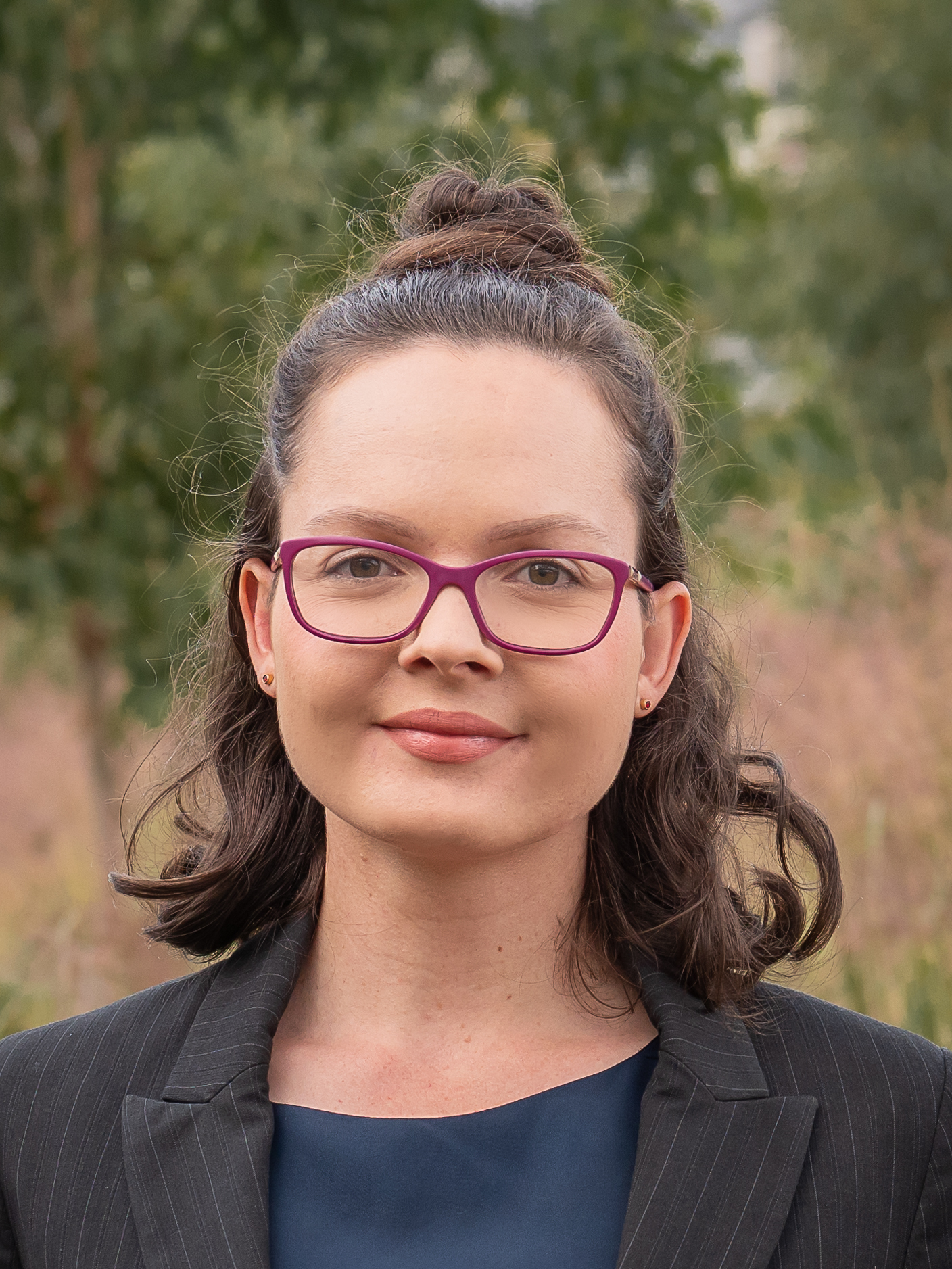
Avrill Aspland
Operations Coordinator
Sydney Cytometry Core Research Facility
The Centenary Institute and the University of SydneyAvrill is the oerations coordinator for Sydney Cytometry, where she is responsible for the management of biosafety processes. She provides key technical expertise for the application of biosafety control measures, collaborating closely with safety officers and researchers. Through this collaboration and oversight of the safety framework she facilitates efficient access to technologies, while maintaining a safe space for all staff and users. Avrill is a member of the ISAC Biosafety Committee and is looking forward to contributing further in this space.
Iyadh Douagi,PhD
Chief, Flow Cytometry Section
Research Technologies Branch
NIAID
NIHDr. Douagi is chief of the Flow Cytometry Section at the Research Technologies Branch, National Institute of Allergy and Infectious Diseases, National Institutes of Health. He received a MS in biochemistry in 1996 from the University of Nice Sophia Antipolis in France, followed by a PhD in immunology in 2001 from Pasteur Institute, Paris. After a postdoctoral training in preclinical discovery at AstraZeneca Sweden, he joined the Swedish Institute for Infectious Disease Control and later Karolinska Institutet in Stockholm as assistant professor to work in vaccine research, exploiting the interplay between innate and adaptive immunity toward viral pathogens including HIV, Rotavirus, and Crimean-Congo hemorrhagic fever virus.
Dr. Douagi has co-authored over 50 peer reviewed articles including publications in National Medicine, Nature Cell Biology, Cell Stem Cell, Nature Communications, and The Journal of Experimental Medicine. His research focuses on providing full stack integration of cytometry-based technologies to investigate cell fate decisions and trajectory inference in health and disease conditions. His contributions include the early development of single-cell technologies to define the repertoire of antigen-specific B and T cells and, recently, the implementation of comprehensive profiling of cell surface proteins by flow cytometry for the identification of novel cell state-specific markers.
Dr. Douagi strongly advocates for the importance of education in support to the next generation of scientists and actively strives to identify opportunities to integrate science and technology with research education. In 2012, he initiated a doctoral course in flow cytometry and regularly participates as faculty in international courses and workshops. Dr. Douagi is former president of the Swedish Society for Flow Cytometry and an active member of the International Society for Advancement of Cytometry, where he serves on the Education and Biosafety Committees.
Stephen Perfetto, MS, MT(ASCP)
Chief, Flow Cytometry Core Section
National Institute of HealthStephen P. Perfetto received both BS degree in medical technology and an MS degree from West Virginia University. During his earlier career, he managed a core flow cytometry facility at the Walter Reed Army Institute of Research where he was involved in large HIV vaccine trials and developed functional tools to study the immune systems of infected individuals. In 2000, he joined the NIH Vaccine Research Center (VRC) as a staff scientist and manager of the flow core facility, a world leader in multicolor flow cytometry with BSL-2 and BSL-2 plus laboratories. His current major areas of research include development, advancement, and application of novel multidimensional and multicolor flow cytometry techniques; detailed assays to an in vivo system to explore the role of CTL in lymphocyte dynamics during viral replication; identification of viral reservoirs in CD4 T-cell subsets; and analysis and sorting of HIV antigen-specific B cells to study neutralizing antibodies and correlate the immunophenotyping of these cells using index sorting.
CMLE Credit: 1.0
-
Contains 3 Component(s), Includes Credits
A CYTO Virtual Interactive 2021 Scientific Tutorial presented by Florian Mair and Thomas Liechti Keywords: immunophenotyping; panel design, experimental design, spreading error, high-parameter cytometry, troubleshooting, best practices
Overview
Modern instrumentation allows immunophenotyping assays to measure up to 40 parameters, which can provide critical insight into the function of the human immune system. However, in order to generate high-quality data without artifacts, these complex assays require thorough experimental planning and robust controls.
In this webinar, we will provide a practical guide for successful experiment design for large immunophenotyping studies using panels from 10-40 (or more) parameters, both for conventional compensation-based as well as spectral cytometry.
Specifically, we will cover the following topics:
- The concept of spreading error (SE) and how to use it for panel design.
- General considerations for setting up large immunophenotyping projects.
- An overview of typical caveats and essential controls.
Speakers
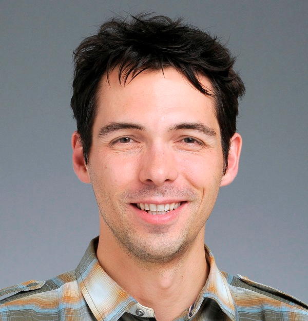
Florian Mair, PhD
Immunologist
Fred Hutchinson Cancer Research CenterFlorian Mair graduated with a PhD from the University of Zurich, Switzerland and is currently working at the Fred Hutchinson Cancer Research Center as an immunologist. During the past decade, he has been involved extensively with different cytometry platforms as well as scRNA-seq techniques and developed an interest in applying novel analysis approaches for single cell data. He has been actively engaged in teaching flow cytometry courses, including systematic panel design and analysis of high-dimensional cytometry experiments. He is also an ISAC Marylou Ingram Scholar.
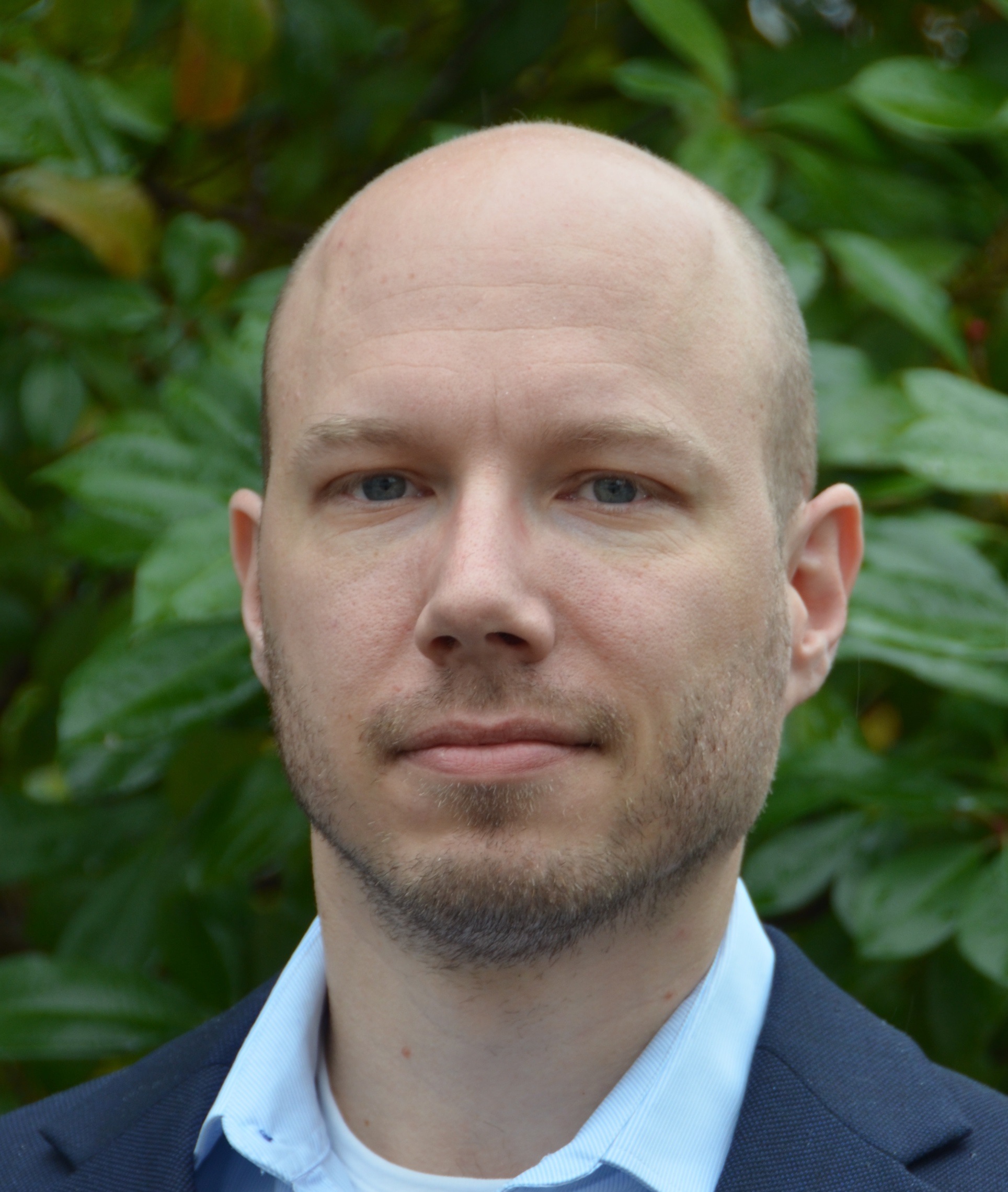
Thomas Liechti, PhD
Postdoctoral Researcher
Vaccine Research Center
National Institute of HealthThomas Liechti obtained his PhD in immunology and microbiology at the University of Zurich and is currently a postdoctoral researcher in Mario Roederer’s group at the Vaccine Research Center of the National Institutes of Health in Bethesda (USA). His main interest is high-dimensional flow cytometry and human immunology. During his postdoctoral training, he established a 28-color flow cytometry sample processing and analysis pipeline to assess the contribution of genetic and environmental factors to human immune homeostasis in a cohort of more than 3,000 individuals.
CMLE Credit: 1.0
-
Contains 3 Component(s), Includes Credits
A CYTO Virtual Interactive 2021 Scientific Tutorial presented by Rob Salomon, David Gallego-Ortega, and Luciano Martellotto Keywords: Genomic cytometry, data analysis, single cell multi-omics, sc-RNAseq, best practices
Overview
Genomic cytometry and the concurrent advancement in associated technologies is fundamentally reshaping our ability to push the dimensionality barrier to answer fundamental questions in biology that were previously impossible. This tutorial will look at the current state of cytometry, discuss how to build complex genomic cytometry pipelines and provide tips and hints on how to match equipment and approaches to the biological question with the view to producing robust, cost-
effective data of the highest quality.Speaker
Rob Salomon
Operations and Technology Manager
ACRF Child Cancer Liquid Biopsy Program
Children's' Cancer InstituteRob is a scientifically trained technologist, internationally recognized for his innovative approach to building large-scale multidisciplinary research programs based around technologically-advanced instrumentation and the broad incorporation of genomics, cytometry, and microfluidics to build high-sensitive multi-omic approaches. Originally trained as cytometrist, Rob now works at the edge of biology and engineering focusing on the application of novel technologies in liquid biopsy to answer clinically-relevant questions in childhood cancer. Having designed, built and run shared resource laboratories of national and international standing, Rob has supported hundreds of research groups and enabled robust data generation for thousands of biological questions.
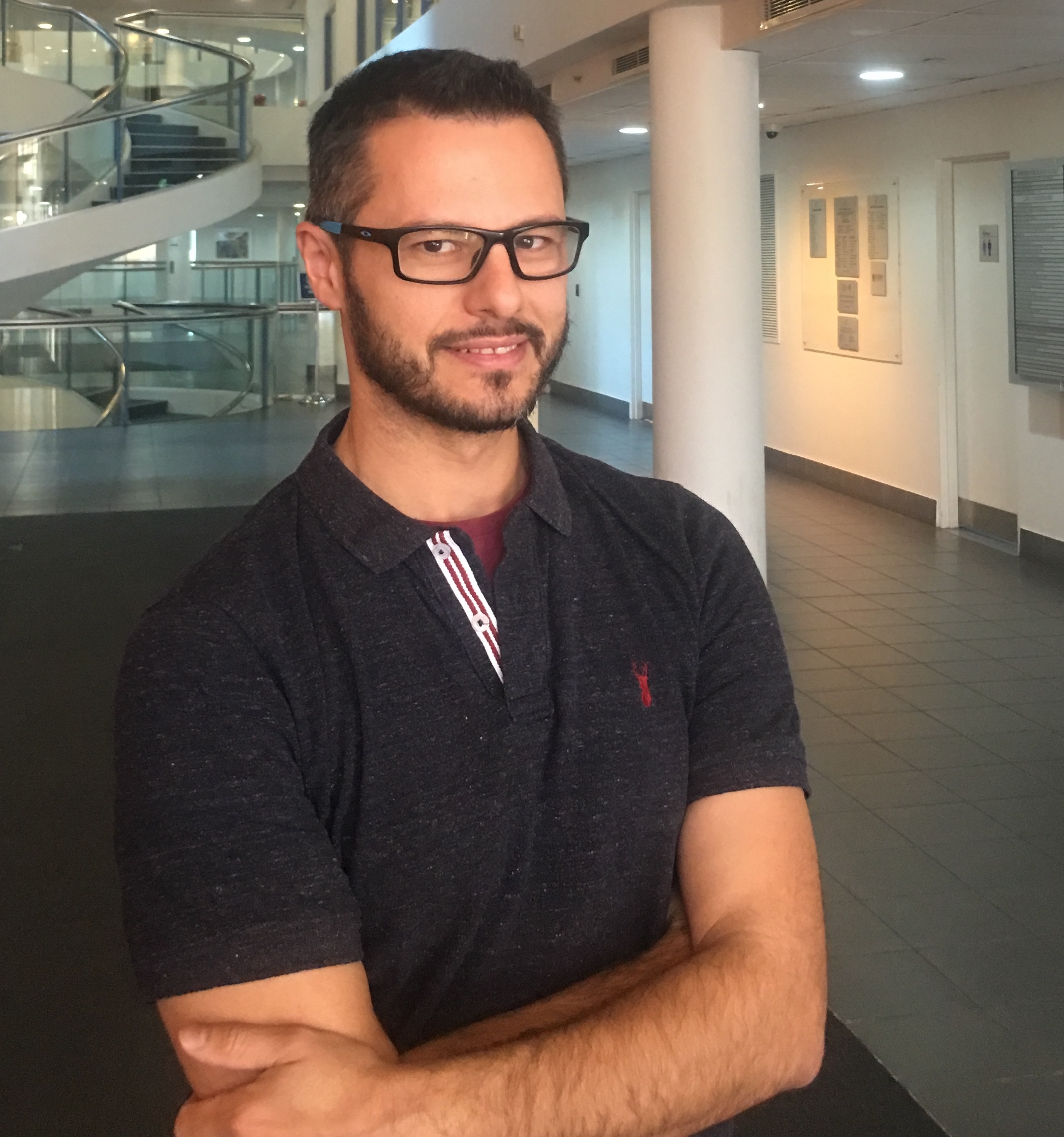
David Gallego-Ortega, PhD
Director
Centre for Single-Cell Technologies
University of Technology SydneyDavid was awarded a PhD in biochemistry, molecular biology and biomedicine in July 2008 at the Universidad Autonoma de Madrid in Spain. After a brief position at TCD Pharma, a biotechnology spin-off company, he moved to the Garvan Institute of Medical Research in 2009 to pursue postdoctoral studies on mammary gland development and breast cancer. David has held an Early Career Fellowship from the National Breast Cancer Foundation and a Career Development Fellowship from the Cancer Institute NSW.
David is currently director of the Centre for Single-Cell Technologies at the University of Technology Sydney, and he holds the Elaine Henry Fellowship from the National Breast Cancer Foundation.
Using a combination of invivo and tissue-engineered ex vivo models, his research group uses single-cell resolution approaches to study developmental mechanisms of the mammary gland and the progression to breast cancer. His research interests focus on the mechanisms of communication between different cellular compartments within the tissue ecosystem and, in particular, inflammatory pathways driven by myeloid cells during mammary morphogenesis and metastatic cancer dissemination.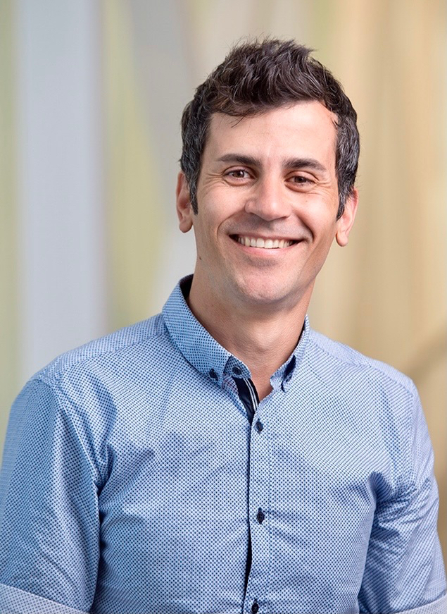
Luciano Martellotto, PhD
Scientific Director
Single Cell Core Laboratory
Harvard Medical SchoolLuciano Martelotto, formerly head of the Single Cell Innovation Lab (SCIL) at the University of Melbourne Centre for Cancer Research (UMCCR), currently holds the full-time position of scientific director of the Single Cell Core Laboratory at Harvard Medical School, in the Department of Systems Biology. Dr. Martelotto has made seminal discoveries in the area of Cancer Biology and Genomics with a focus on single cell analysis of biological systems. As a Research fellow at the Memorial Sloan Kettering Cancer Centre (USA) he co-authored more than 30 articles in 3 years, including first authorships in the top-tier journals Nature Medicine, Nature Genetics and Genome Biology. Dr. Martelotto has published more than 50 articles in diverse areas of science, including first authorships in top-tier journals like Nature Medicine (2011, 2017a, 2017b, FWCI), Nature Genetics (2014, FWCI), Genome Biology (2014a, 2015b, FWCI) and others. Martelotto holds patent inventorships in Europe and the US. Dr. Martelotto is currently a scientific advisor at OmniScope. From 2017 to 2020 he ran the new SCIL at the UMCCR where he established a functional a state-of-the-art Single Cell Laboratory that became the go-to lab to set out new and challenging projects both inter-state and internationally. This success led to his recruitment as Scientific Director of the Single Cell Core Laboratory at Harvard Medical School. Here, Luciano leads all the scientific aspects and collaborative efforts of the Single Cell Core Laboratory at Harvard Medical School. Dr. Martelotto and his team oversee the implementation and development new technologies in the field of single cell genomics and spatial transcriptomics and makes them available to the wider Harvard Community. Dr. Martelotto has a large scientific network and maintains strong collaborative ties with the Australian Scientific community.
CMLE Credit: 1.0

