Fluorescence Cytometry
-
Register
- Visitor - $200
- Bronze Member - Free!
- Platinum Member - Free!
- Platinum - Free!
- Bronze Member 3-Year - Free!
- Silver Member 3-Year - Free!
- Platinum Member 3-Year - Free!
- Single Cell Analysis of Autofluorescence Lifetime Images
- Multiplexed Fluorescence Microscopy Reveals Heterogeneity among Stromal Cells in Mouse Bone Marrow Sections
- Evaluating Spectral Cytometry for Immune Profiling in Viral Disease
- The Essentials of Optics and Fluorescence Microscopy for Cell Biology Applications (2013 Advanced Data Analysis Pre-Congress Course)
-
Contains 3 Component(s), Includes Credits Recorded On: 11/19/2019
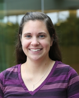
A CYTO U Webinar presented by Alex Walsh, PhD Keywords: Optical imaging, 2-photon imaging, cancer therapy, Autofluorescence
About the Presenter

Alex Walsh, PhD
Assistant Professor in Biomedical Engineering
Texas A&M University
Dr. Walsh completed her Ph.D. at Vanderbilt University, where she developed an autofluorescence lifetime-based assay for determining the optimal cancer treatment strategy for individual patients. As a postdoc at the Air Force Research Lab, Dr. Walsh used optical techniques to investigate infrared-light activation and inhibition of action potential propagation in neurons. Currently, Dr. Walsh is an assistant professor in the Biomedical Engineering Department at Texas A&M University.Webinar Summary
Fluorescence lifetime imaging (FLIM) of the endogenous fluorophores, NAD(P)H and FAD (co-enzymes of metabolic reactions), provides a label-free method to quantify cellular metabolism. This webinar will review multi-photon fluorescence lifetime imaging methods, single cell segmentation, and intra-population heterogeneity analysis. Examples will be shown for drug response in breast cancer organoids and activation of T cells.
Learning Objectives
- Define fluorescence lifetime and time-correlated single photon counting imaging methods.
Interpret label-free FLIM images of NAD(P)H and FAD. - Discuss segmentation and single-cell analysis techniques.
Who Should Attend
Anyone interested in label-free imaging, fluorescence lifetime imaging, or single-cell analysis.
CMLE Credit: 1.0
- Define fluorescence lifetime and time-correlated single photon counting imaging methods.
-
Contains 3 Component(s), Includes Credits Recorded On: 04/15/2019
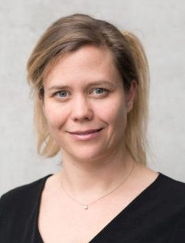
A CYTO U Webinar presented by Anja E. Hauser, PhD Keywords: multiplex histology, quantitative, bone marrow
About the Presenter

Anja Hauser, PhD
Professor of Immune Dynamics and Intravital Microscopy
Charité—Universitätsmedizin and Deutsches RheumaforschungszentrumDuring her studies of veterinary medicine, Anja Hauser developed an interest in immunopathology and microscopy. As a graduate student, she identified factors which attract plasma blasts into the bone marrow and keep them alive in specialized niches within this tissue. During her postdoctoral work, she worked on the migration of germinal center B cells using intravital 2-photon microscopy. She is a professor of immune dynamics and intravital microscopy at the Charité—Universitätsmedizin and Deutsches Rheumaforschungszentrum in Berlin. In an interdisciplinary approach, her lab develops novel microscopy technologies to obtain a deeper insight in how the immune system functions.
Webinar Summary
Anja will introduce the principles of multi-epitope ligand cartography (MELC), a method for multiplexed immunofluorescence microscopy, and explain its application for analyzing complex tissues and rare cell subsets. She will also give an overview on methods suitable to quantitatively analyze those complex multi-parametric image data.
Learning Objectives
- Describe MELC as a method for multiplexed immunofluorescence histology.
- Learn what to consider when preparing tissues for MELC analysis, such as choosing marker panels and planning the sequential staining in the tissue
- Learn about options for image analysis in order to extract quantitative information from multiplexed immunofluorescence histology.
Who Should Attend
Everyone who is interested in quantitative multiplexed image analysis and its applications in immunology.
CMLE Credit: 1.0
-
Contains 4 Component(s), Includes Credits Recorded On: 05/18/2013
A CYTO 2013 Introduction to Image-Based Cytometry PreCongress Course presented by Stephen J. Lockett Keywords: basics of imaging, microscopy, CYTO 2013
The Presenter
Stephen Lockett
Optical Microscopy and Analysis Laboratory
Frederick National Laboratory
National Cancer Institute
Frederick, MD 21702Session Summary
Modern optical microscopy techniques have enabled the acquisition of quantitative information from cells with unprecedented accuracy and specificity. This technology is poised to continue making a significant impact in health-related sciences by facilitating diagnosis as well as enabling important discoveries related to cellular mechanisms. The purpose of this short course is to enable scientists and technical practitioners to utilize the most commonly available optical imaging hardware and software tools to quantitatively describe and analyze cellular processes. It aims to provide the necessary background as well as to introduce the terminology, concepts, and methodological approaches employed in modern image cytometry and high-content screening. The course will expand and strengthen the participants' knowledge and understanding of modern digital high-content microscopy techniques. It will provide participants with the required tools for quantitative interpretation of image-based experiments.
The course is intended for researchers and students who have previous experience with other optical methods such as flow cytometry or basic optical microscopy.
Topics covered in this session include:
- Image formation (diffraction, magnification, etc.).
- Fluorescence (staining, photodamage, etc.).
- 3D and live cell imaging.
CMLE Credit: 1.0
-
Contains 3 Component(s), Includes Credits
A CYTO U Webinar presented by Paula Niewold, PhD and Thomas Ashhurst, PhD Keywords: spectral cytometry, unmixing, compensation, conventional cytometry, autofluorescence, panel design, spreading error, platform comparison, data analysis
About the Presenters

Paula Niewold, PhD
Postdoctoral Researcher
Department of Infectious Diseases
Leiden University Medical CentreDr. Paula Niewold is an immunologist currently working as a postdoctoral researcher at the Department of Infectious Diseases at the Leiden University Medical Centre. She is interested in host-pathogen interactions and how they impact the outcome of disease. She has studied these interactions in models of cerebral malaria, West Nile virus encephalitis, psoriasis, and tuberculosis using high-dimensional flow, mass, and imaging mass cytometry. She is an ISAC Marylou Ingram Scholar.

Thomas Ashhurst, PhD
Immunologist and High-Dimensional Cytometry Specialist
Sydney Cytometry Facility
University of SydneyDr. Thomas Ashhurst is an immunologist and high-dimensional cytometry specialist with the Sydney Cytometry Facility at the University of Sydney. He develops and applies a range of single-cell cytometry technologies and computational analysis tools to map dynamic immune responses over time, space, and disease. In particular, he applies these approaches to the study of immunology and infectious disease, including emerging pathogens such as COVID-19, Zika virus encephalitis, and West Nile virus encephalitis. He is an ISAC Marylou Ingram Scholar.
Webinar Summary
In conventional fluorescence cytometry, each fluorophore in a panel is measured in a target detector, through the use of wide band-pass optical filters. In contrast, spectral cytometry uses a large number of detectors with narrow band-pass filters to measure a fluorophore's signal across the spectrum, creating a more detailed fluorescent signature for each fluorophore. The spectral approach shows promise in adding flexibility to panel design and improving the measurement of fluorescent signal. However, few comparisons between conventional and spectral systems have been reported to date. Here we present our findings comparing conventional and spectral approaches to cytometry—including comparisons of compensation and unmixing—and evaluate the use of spectral cytometry for immune profiling in viral diseases.
Learning Objectives
- Gain an understanding of the essential differences between conventional and spectral approaches to cytometry.
- Appreciate the differences between compensation and spectral unmixing.
- Consider applications for spectral cytometry in the context of immunological studies.
Who Should Attend
Researchers and technical staff who utilize flow, spectral, or mass cytometry in their work.
CMLE Credit: 1.0
-
Contains 4 Component(s), Includes Credits Recorded On: 10/21/2020
A CYTO U Webinar presented by Florian Mair, PhD Keywords: panel design, antibody titration, compensation, thawing, PBMCs
About the Presenter

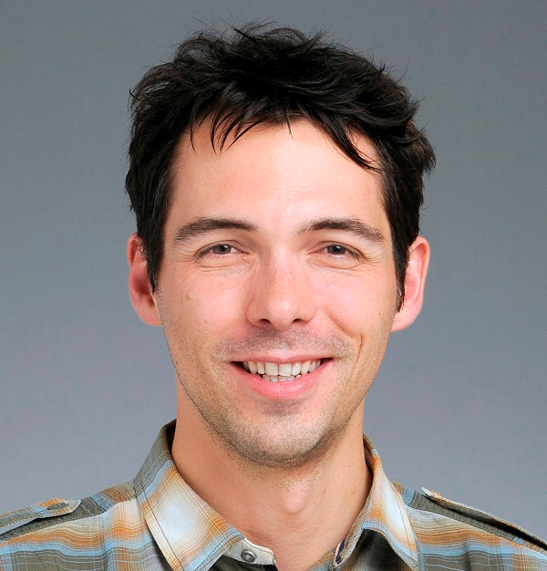
Florian Mair
Cytometry Specialist
Fred Hutchinson Cancer Research CenterFlorian graduated with a PhD from the University of Zurich, Switzerland in 2014 and is currently working at the Fred Hutchinson Cancer Research Center as an immunologist. During the past decade, he has been involved extensively with different cytometry platforms (conventional, spectral, and mass cytometry) as well as scRNA-seq technique. Florian is interested in applying novel analysis approaches for single cell data. He has been actively engaged in teaching flow cytometry courses including systematic panel design and analysis of high-dimensional cytometry experiments. Florian is currently an ISAC Marylou Scholar.
Webinar Summary
Over the past decade, technical improvements and new reagents have permitted fluorescent-based flow cytometry assays to measure up to 40 parameters. These complex assays require robust controls and thorough experimental planning, but there are currently few resources that provide a systematic approach for reliable panel design. Also, historical notions as to how fluorophores and controls should be chosen are sometimes at odds with the reality of modern panel design.
In this webinar, we will provide a practical guide for successful fluorescent panel design for any complex panel from 10–40 (or more) parameters, both for conventional compensation-based as well as spectral cytometry.
Specifically, we will cover the following topics:
- Brief overview of signal detection in conventional and spectral flow cytometers.
- The concept and underlying cause of spreading error (SE).
- How the spillover spreading matrix (SSM) can be efficiently used to guide panel design.
- Relevant relationships between SE, fluorophore brightness, and antigen expression level.
- Step-by-step approaches toward building a new panel.
- An overview of essential controls and typical caveats.
This webinar is back-to-back with a webinar by Thomas Liechti, who will be talking about how to best design and use complex panels for large study cohorts (100s-1000s of samples) including the use of appropriate controls and analysis approaches.
Learning Objectives
- An understanding for tackling high-dimensional immunophenotyping assays on a variety of platforms, thus minimizing trial-and-error experiences and wasted experiments
- Review specific strategies for generating reproducible and high-quality data in large patient cohorts.
Who Should Attend
Anyone with an interest in efficient panel design: immunologists, scientists of any field doing polychromatic flow cytometry, SRL users, and SRL leaders.
CMLE Credit: 1.0

