
Cytometry Panels
-
Register
- Visitor - $50
- Bronze Member - Free!
- Platinum Member - Free!
- Platinum - Free!
- Bronze Member 3-Year - Free!
- Silver Member 3-Year - Free!
- Platinum Member 3-Year - Free!
- The Road to a High-Resolution 40-Color Flow Cytometry Immunophenotyping Panel
- Designing Panels for the Study of Hematopoietic Stem Cells
- Panel Design - A Practical Guide for Successful Fluorescent Cytometry Panels from 10-40 Parameters
- Predicting the Best Resolution and Sensitivity in Panel Development and Reducing Inter-instrument Variability in Flow Cytometry
- 16-Color Panel to Measure Inhibitory Receptor Signatures from Multiple Human Immune Cell Subsets
- Step-by-Step Multi-parameter Panel Design
-
Contains 4 Component(s), Includes Credits Recorded On: 10/15/2018
A CYTO U Webinar presented by Jennifer Wilshire, PhD & Tomas Baumgartner Keywords: panel design, spillover spreading, spillover spreading matrix, resolution impact matrix, sentinel panel design, brightness, compensation, controls, spectra
The Presenters

Jennifer Wilshire, PhD
Assistant Manager
Memorial Sloan Kettering Cancer Center
Tomas Baumgartner
Manager
Flow Cytometry Core Facility
Weill Cornell MedicineWebinar Description
This webinar will focus on the process of building multicolor panels. We will break the process down into step-by-step guides based on the number of colors in your panel. This webinar will also provide a framework for those teaching multicolor panel design in an educational setting such as a Shared Resource Lab.
Learning Objectives
- Know the three processes used to build multicolor panels.
- Choose the correct step-by-step process based on the number of colors in the panel.
- Interpret spillover spreading matrix/resolution impact matrix to determine which colors contribute spreading to others.
- Practice building a multicolor panel using all the tricks and tips discussed in the webinar.
Who Should Attend
Anyone who is designing multicolor panels or teaching others how to design multiparameter panels.
CMLE Credit: 1.0
-
Contains 4 Component(s), Includes Credits Recorded On: 08/09/2018
A Cytometry Part A Spotlight CYTO U Webinar presented by Anna Belkina, MD, PhD Keywords: Panel design, optimization, high-parameter, OMIP, exhaustion
About the Presenter

Anna Belkina, MD, PhD
Associate Director of the Flow Cytometry Core Facility
Assistant Professor of Pathology and Laboratory Medicine
Boston University School of MedicineAnna Belkina is the associate director of the Flow Cytometry Core Facility and an assistant professor of pathology and laboratory medicine at Boston University School of Medicine. She received her MD from Russian State Medical University in Moscow and her PhD degree from Boston University School of Medicine investigating the epigenetic regulation of inflammatory responses driven by bromodomain proteins. Anna’s research is focused on the intersection of immunology and computational biology. Her current efforts include investigating the immune landscape of chronic inflammatory diseases and developing computational techniques to assess high-parameter single cell cytometry data. Anna is an active member of ISAC and was named an ISAC SRL Emerging Leader in 2015.
Webinar Summary
This webinar will walk you through the development and optimization of a 16-color human flow cytometry panel for combinational analysis of the Inhibitory Receptors PD-1, TIGIT, CD160, LAG-3, TIM-3 and the activation marker CD137 from CD4+ T cells, CD8+ T cells, NK cells, iNKT cells, and gamma delta T cells. A flow cytometry panel that measures 5-plex IR signatures from multiple populations of immune cells would be useful for both mechanistic studies of exhaustion as well as translational research efforts for a wide span of chronic diseases such as cancer and HIV, especially in the context of scarce sample material.
This panel was thoroughly optimized for use on a 4-laser, 16-detector BD FACSARIA II SORP. In order to gain optimal performance from using multiple polymer fluorophores, panel design was preceded by instrument calibration and optimization based on previous publications. The selection of reagents was defined by predicted marker distribution on cells and levels of their expression and fluorophore "brightness," as well as predicted spillover spread within the fluorophore matrix and reagent availability from common suppliers. When multiple clones were available for particular antigens of interest, selections were made after thorough literature review. Finally, FMO tests were performed to verify specificity of observed populations. Several reagents to limit spillover spread in the channels that appeared to be more vulnerable to this issue.
The conclusion will include some data from a large-scale study, where this panel has been employed, to successfully map an IR-specific phenotype in a cohort of aviremic HIV+ individuals and link it to disease.
Learning Objectives
- Recall basic principles of high-parameter panel design and prerequisite instrument optimization.
- Review major human immune subset mapping with surface markers.
- Demonstrate the process of panel development and optimization.
- Evaluate the resulting panel design with real life application.
Who Should Attend
High-parameter flow cytometry practitioners including primary researchers and core facility staff, as wellas scientists who want to start building their own extensive multicolor flow cytometry panels.
CMLE Credit: 1.0
-
Contains 4 Component(s), Includes Credits Recorded On: 04/28/2015
CYTO U Webinar presented by Steve Perfetto and Jim Wood, PhD Keywords: linearity, reproducibility, calibration, noise, resolution, precision, Pulsed LED, Q&B
About the Presenters

Stephen Perfetto
Chief, Flow Cytometry Core Section
NIHStephen Perfetto received a BS in medical technology from the West Virginia University in 1977 and completed his MS in 1981 from West Virginia University. He studied and worked in the clinical blood bank and clinical immunology laboratories until 1988, when he joined the EPICS Division of Coulter Corporation. In 1990 he was recruited to the Walter Reed Army Institute of Research and was the manager of the core flow facility. While at WRAIR, he was involved in large HIV vaccine trials and developed functional to study the immune system of infected individuals.
In 2000 he joined the NIH in the department of the Vaccine Research Center (VRC) as a staff scientist and manager of the flow core facility. This facility is the world leader in multicolor flow cytometry and continues to actively develop this technology on a number of different fronts. One focus is on hardware development and reagent and analysis development. For several years, this lab has collaborated to develop Quantum Dots for use in immunophenotyping experiments.

James Wood, PhD
Flow Cytometry ConsultantJames Wood obtained his BA in physics from Gettysburg College; however, he also availed himself to a broad range of general and advanced biology and chemistry courses during his undergraduate years. He completed his MS and PhD degrees in biophysics from The Pennsylvania State University studying the effects on the life cycle of cells after radiation exposure. In graduate school, he also developed the first microprocessor-based two-parameter flow cytometer data acquisition system. After completing a postgraduate position in the lab of Dr. Leon Wheeless at the University of Rochester Medical Center where he contributed to the development of a 3D slit-scan flow cytometer, he moved to Florida accepting a position with the EPICS Division of Coulter Corporation. He was manager of the New Products Research and Applications Laboratory during most of his tenure at Coulter and Beckman-Coulter. He currently consults for flow cytometry and pharmaceutical companies and manages the flow cytometry shared resource at Wake Forest University School of Medicine Comprehensive Cancer Center (WFUCCC).
Webinar Summary
Successful quantitative flow cytometry requires an understanding of the characteristics of a flow cytometer instrument that affect the measurement and analysis of the acquired photometric data. Flow cytometer instruments must be characterized before being used for critical work. The immediate goals include achieving better reproducibility of data, reducing variation and to facilitate intra/inter-lab comparisons of cytometry data. Characterization includes the determination of the instrument linearity, dynamic range, precision, accuracy, detection efficiency (Q), and electronic and optical noise (B). In particular, Q and B affect how well cellular receptors with low expression can be measured. For example, staining panels may be inadvertently designed to avoid measuring dim markers on PMTs with poor resolution. Unfortunately, current methods using bead sets to assess PMT resolution only provide rough estimates of Q and B. Thus, staining panel design and similar protocols remain a largely empirical process, requiring time and intimate experience with an instrument’s performance. Ascertaining these instrument characteristics are the first steps toward assessing how instruments can be standardized and calibrated. Ultimately, this may be part of the validation process to certify that a flow cytometer used for critical clinical and/or GMP work is performing at the required level needed to obtain accurate and precise results. Additionally, as part of this process we need to move from the common use of “relative intensity units (channel numbers)” to data measured and presented in physically relevant optical units (photons, photoelectrons, dye molecules, antibody binding sites).
Recently, James Wood (Wake Forest University) developed a new device, known as an LED Pulser, to measure B and Q directly and accurately. This device delivers consistent, uniform broad-spectrum light pulses, the amplitude of which can be adjusted with an electronic and/or an optical attenuator. Our presentation will demonstrate how this device is used to calculate Q and B values. In the past, data from multilevel bead sets have been used to facilitate comparisons of instrument sensitivity with Q and B; however, it has proved difficult to manufacture bead sets that have closely matched intrinsic CVs. In practice the LED Pulser, along with calibrated fluorochrome-loaded beads, can be used to determine the relationship between fluorescence channel numbers and the number of photoelectrons generated at the PMT photocathode (i.e., the statistical photoelectron estimate (Spe) using a weighted quadratic fitting of pulsed LED series data). This information, along with estimates of receptor density, can be used to calculate index values that provide quantitative guidance for panel design. Our presentation will describe this process step-by-step with examples. Additionally, we will show how the metrics can be used for inter-laboratory comparisons.
The first part of the webinar will present the principles behind the LED Pulser, as well as the appropriate use of the LED Pulser and how it compares to using multilevel bead sets for the calculation of Q and B. The second part of the webinar will introduce how the LED Pulser, along with calibrated fluorochrome-loaded beads lead to better estimates of the four facets of predictive panel design: (i) accurate Q and B values for each detector, (ii) optimal voltage settings, (iii) spillover/spreading error calculations, and (iv) estimates of receptor density and contribution to more quantitative determinations for optimizing panel design.
CMLE Credit: 1.0
-
Contains 4 Component(s), Includes Credits Recorded On: 10/29/2020
A CYTO U Webinar presented by Dr. Steffen Schmitt and Dr. Marcus Eich Keywords: panel design, OMIP, history, autofluorescence, antibody titration
About the Presenters

Dr. Steffen Schmitt
German Cancer Research Center (DKFZ)Steffen Schmitt studied biology at the Ruprecht-Karls-University Heidelberg and finished his PhD with a concentration in immunology. Subsequent to his postdoc, he developed the flow cytometric service at the Center for Natural and Medical Sciences (NMFZ) at Johannes-Gutenberg-University in Mainz. Since 2007 Steffen has headed the Flow Cytometry Core Facility of the German Cancer Research Center (DKFZ) in Heidelberg.

Dr. Marcus Eich
Hi-Stem gGmbHMarcus Eich studied biology at the Technical University Darmstadt and earned his PhD in 2011 in toxicology at the University Medical Center Mainz. After an additional postdoc in Mainz in 2015, he joined the HI-STEM team and the Flow Cytometry Core Facility of the German Cancer Research Center (DKFZ) in Heidelberg.
Webinar Summary
The hematopoietic system is a very fascinating system to study as it can re-populate an entire system starting from just one cell. This webinar gives an overview of the targeted subpopulations of the hematopoietic system and how different experimental tasks and setups in one panel were combined. At the end, issues that occurred during the panel design are discussed and a short outlook is presented about how to further improve the panel.
Learning Objectives
- Understand the hematopoietic system from a backbone panel to specialized subpopulations, combining different experimental setups in panel design.
- Apply tips and tricks for this panel.
Who Should Attend
PhD students, postdocs, and technicians involved in hematopoietic stem cell research.
CMLE Credit: 1.0
-
Contains 3 Component(s), Includes Credits
A CYTO U Webinar presented by Maria C. Jaimes, MD Keywords: High-dimensional flow cytometry, spectral flow cytometry, OMIP, Panel development
About the Presenter
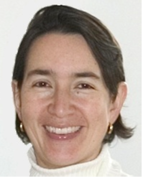
Maria C. Jaimes, PhD
VP Technical Application Support
Cytek Biosciences, Inc.Dr. Maria Jaimes earned her MD degree at the Universidad Javeriana in Colombia. Dr. Jaimes completed her postdoctoral training at Stanford University in the Department of Microbiology and Immunology. During her postdoc, she focused on characterizing the immune responses to both rotavirus and influenza viruses after natural infection and immunization. In 2005, Dr. Jaimes joined BD Biosciences, where she worked on different aspects of quality assurance and standardization of flow cytometry assays. In 2015, Dr. Jaimes joined Cytek Biosciences. She is part of the R&D team credited for developing the Aurora Full Spectrum Cytometer and has overseen the instrument characterization, verification, and development of multicolor applications. Besides her responsibilities within the R&D team, Dr. Jaimes leads the Technical Applications Support team worldwide.
Webinar Summary
This webinar will cover OMIP-069 (published in Cytometry Part A in August 2020), the first 40-color fluorescent panel using full spectrum flow cytometry to broadly phenotype much of the cellular composition of the human peripheral immune system. The panel in this OMIP has been thoroughly optimized to ensure high-quality data and well-resolved populations, enabling the description of most canonical subsets of T cells, B cells, NK cells, monocytes, and dendritic cells. Dr. Jaimes will present the journey of the technology, panel design, protocol development, data QC/QA, and data analysis which led to the successful achievement of this 40-color immune profiling panel.
Learning Objectives
- Understand the concepts behind full spectrum profiling.
- Learn the necessary steps and tools for good panel design.
- Develop expertise on how to troubleshoot and optimize a multicolor panel.
- Learn to recognize high quality vs. compromised data and the potential sources and mitigation of errors.
Who Should Attend
SRL staff/directors, immunologists, CRO staff, and full spectrum flow cytometer users.
CMLE Credit: 1.0
-
Contains 4 Component(s), Includes Credits Recorded On: 10/21/2020
A CYTO U Webinar presented by Florian Mair, PhD Keywords: panel design, antibody titration, compensation, thawing, PBMCs
About the Presenter
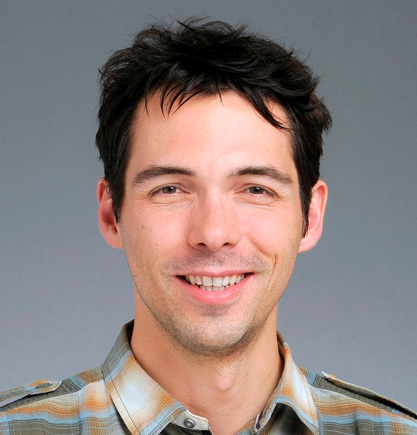
Florian Mair
Cytometry Specialist
Fred Hutchinson Cancer Research CenterFlorian graduated with a PhD from the University of Zurich, Switzerland in 2014 and is currently working at the Fred Hutchinson Cancer Research Center as an immunologist. During the past decade, he has been involved extensively with different cytometry platforms (conventional, spectral, and mass cytometry) as well as scRNA-seq technique. Florian is interested in applying novel analysis approaches for single cell data. He has been actively engaged in teaching flow cytometry courses including systematic panel design and analysis of high-dimensional cytometry experiments. Florian is currently an ISAC Marylou Scholar.
Webinar Summary
Over the past decade, technical improvements and new reagents have permitted fluorescent-based flow cytometry assays to measure up to 40 parameters. These complex assays require robust controls and thorough experimental planning, but there are currently few resources that provide a systematic approach for reliable panel design. Also, historical notions as to how fluorophores and controls should be chosen are sometimes at odds with the reality of modern panel design.
In this webinar, we will provide a practical guide for successful fluorescent panel design for any complex panel from 10–40 (or more) parameters, both for conventional compensation-based as well as spectral cytometry.
Specifically, we will cover the following topics:
- Brief overview of signal detection in conventional and spectral flow cytometers.
- The concept and underlying cause of spreading error (SE).
- How the spillover spreading matrix (SSM) can be efficiently used to guide panel design.
- Relevant relationships between SE, fluorophore brightness, and antigen expression level.
- Step-by-step approaches toward building a new panel.
- An overview of essential controls and typical caveats.
This webinar is back-to-back with a webinar by Thomas Liechti, who will be talking about how to best design and use complex panels for large study cohorts (100s-1000s of samples) including the use of appropriate controls and analysis approaches.
Learning Objectives
- An understanding for tackling high-dimensional immunophenotyping assays on a variety of platforms, thus minimizing trial-and-error experiences and wasted experiments
- Review specific strategies for generating reproducible and high-quality data in large patient cohorts.
Who Should Attend
Anyone with an interest in efficient panel design: immunologists, scientists of any field doing polychromatic flow cytometry, SRL users, and SRL leaders.
CMLE Credit: 1.0

